MRI for Africa: The design of an MRI for the diagnosis of infant hydrocephalus in Ugandan hospitals
Apr 2020 - ongoing
Master thesis by Frank van Doesum
Watch the video to learn about the project.
Magnetic resonance imaging (MRI) is a medical diagnostic imaging technology characterized by its high cost and infrastructure demands. This makes it unsuitable for implementation in low- and medium-income countries. Its costs are directly related to the strength of the system’s magnetic field. Therefore, an MRI scanning technology dedicated for use in low- and medium-income countries has been developed by Delft University of Technology and Leiden University Medical Center that makes use of a low magnetic field. This low-field technology, that requires a smaller size magnet and yields lower resolution images, is already capable of diagnosing infant hydrocephalus, a frequently occurring condition in sub-Saharan Africa. For a successful implementation, it is crucial that this technology is integrated in an MRI system that adequately addresses challenges within the context of sub-Saharan Africa and is equipped with all necessary functionality to diagnose infants with hydrocephalus. From observations and interviews in the healthcare environment of Uganda, it was found that – in addition to high cost – poor infrastructure and challenges that occur during imaging procedures limit the implementation- and use-potential of current MRI systems and thus can affect the implementation of this new system. Poor pathways and unavailability of forklifts or cranes often cause breakage during installation and transport. Moreover, lack of space in hospitals hinders implementation of large systems that require dedicated, shielded rooms. During imaging, movement of the (infant) patient may affect the image, and often mothers are kept in the imaging room with the patient to calm her. Other challenges for the operation of these systems in this context include heat, humidity, dust, power outages and the presence of pests. A concept of a complete MRI system that integrates the developed technology and addresses these challenges is designed.

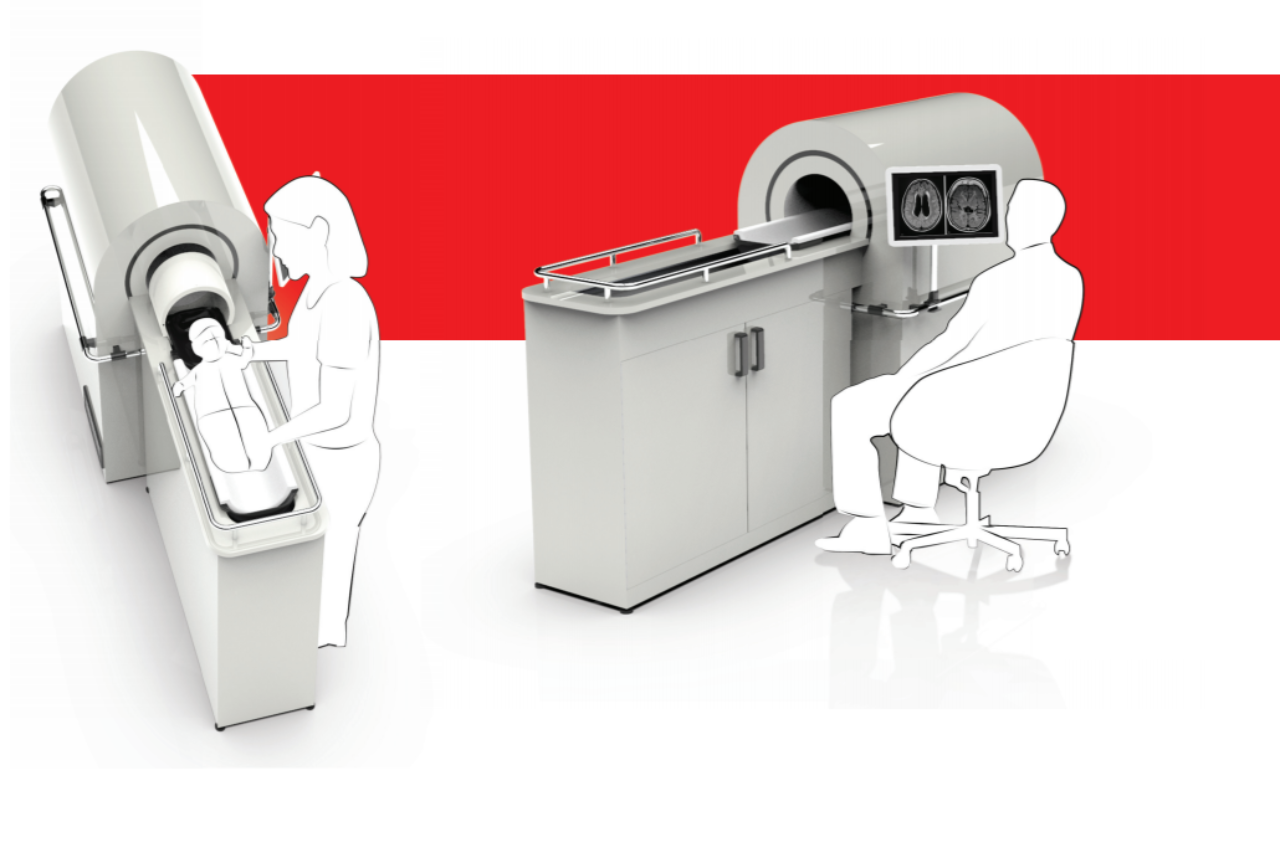
During imaging, movement of the (infant) patient may affect the image, and often mothers are kept in the imaging room with the patient to calm her. Other challenges for the operation of these systems in this context include heat, humidity, dust, power outages and the presence of pests. A concept of a complete MRI system that integrates the developed technology and addresses these challenges is designed. This MRI system enables an ergonomic and safe operation, facilitates safe and comfortable insertion and removal of the patient from the machine, and allows mothers and operators to remain close and in visual contact with the patient. The entire concept takes up no more than 3 m2, can be installed in any room with a power socket and features a plug and play installation. It can be carried over rough pathways without the need for a forklift or crane. Furthermore, the concept features a bed with sliding mechanism that optimizes the space inside the system, allowing hydrocephalus patients with significant expansion of the head to be imaged. With this concept, the first steps are taken towards implementation of the technology in hospitals in low- and medium-income countries. Through further development and testing in close collaboration with stakeholders, the concept can be optimized to enable diagnosis and treatment of infants with hydrocephalus and eventually many others, and hereby contribute to increasing the (e)quality of healthcare around the world.
Read the full graduation report here.